Skip to content
Research

Research & publications

How do motor proteins fold metres of DNA into chromosomes? I study the SMC complexes (condensin, cohesin, Smc5/6 and the bacterial Wadjet) that extrude DNA into loops, by watching them work one molecule at a time. Before that, I used single-molecule fluorescence and gold nanorod antennas to follow electron transfer in single copper proteins.

§ 01

Highlights

Fig. 1Loop extrusion on plasmid DNA. Top, DNA; bottom, DNA (cyan) with JetABCD (red). Scale bar 2 µm.
Molecular Cell 85(1), 107–116 · 2025

Loop-extrusion-mediated plasmid DNA cleavage by the bacterial SMC Wadjet complex

Biswajit Pradhan, Amar Deep, Jessica König, Martin D. Baaske, Kevin D. Corbett, Eugene Kim

Bacteria defend themselves against invading plasmids with an SMC protein machine called Wadjet. Watching single Wadjet complexes at work, we found that they reel a plasmid's DNA into a loop from both sides at once, stall once the whole 44-kilobase plasmid has been pulled in, and then cut it. Loop extrusion is how Wadjet tells a plasmid apart and destroys it.

Abstract

Structural maintenance of chromosomes (SMC) complexes play pivotal roles in genome organization and maintenance across all domains of life. In prokaryotes, SMC-family Wadjet complexes structurally resemble the widespread MukBEF but serve a defensive role by inhibiting plasmid transformation. We previously showed that Wadjet specifically cleaves plasmid DNA; however, the molecular mechanism underlying plasmid recognition remains unclear. Here, we use in vitro single-molecule imaging to directly visualize DNA loop extrusion and plasmid cleavage by Wadjet. We find that Wadjet is a symmetric loop extruder that simultaneously reels in DNA from both sides of a loop and that this activity requires a dimeric JetABC supercomplex. On surface-anchored plasmid DNAs, Wadjet extrudes the full length of a 44-kb-pair plasmid, stalls, and cleaves DNA. Our findings reveal the role of loop extrusion in the specific recognition and elimination of plasmids by Wadjet and establish loop extrusion as an evolutionarily conserved mechanism among SMC complexes across all kingdoms of life.

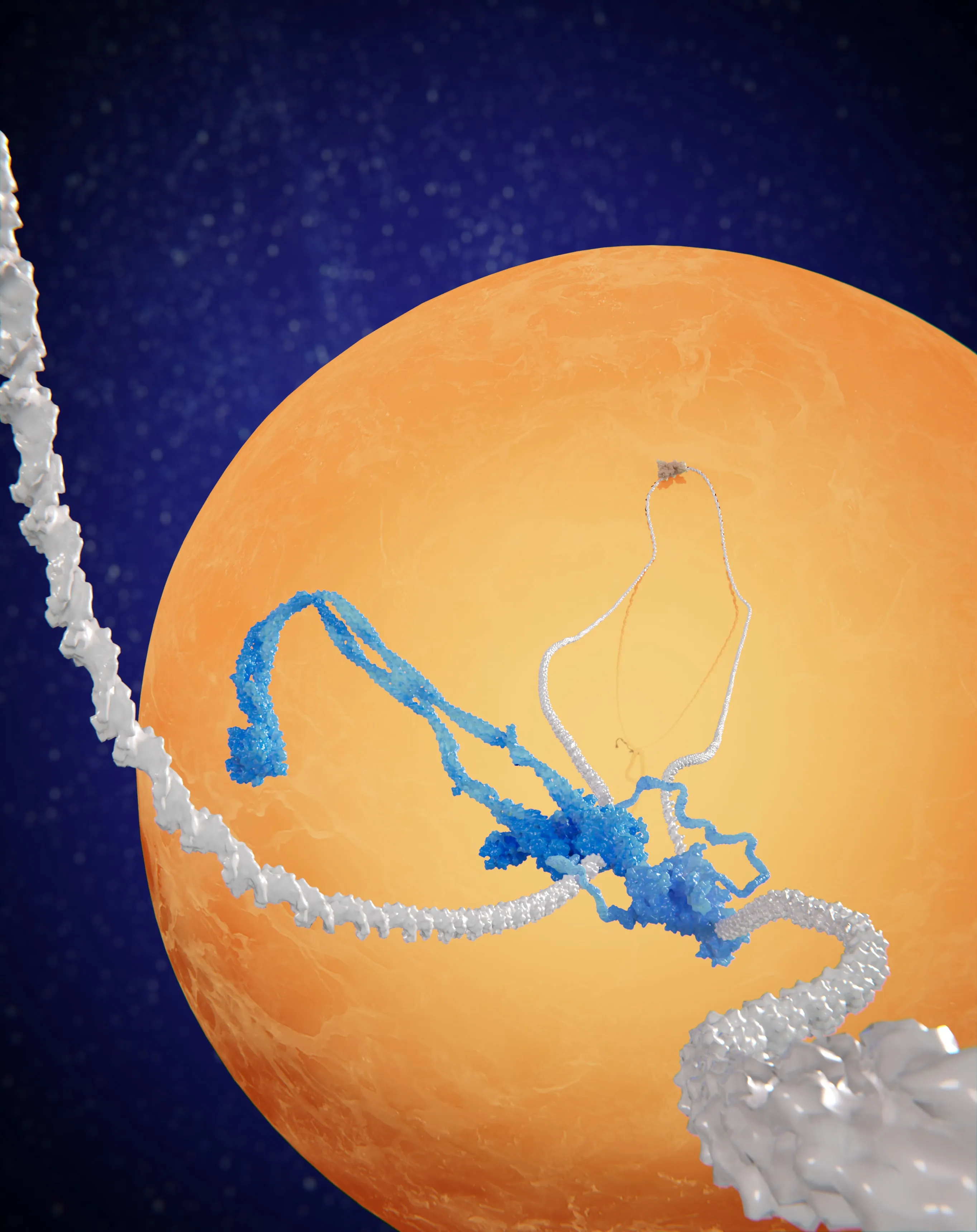
Molecular Cell cover, volume 84 issue 5 — a pearl necklace twisted into a looped, supercoiled knot among jewellery
Fig. 2Cover of Molecular Cell 84(5), March 2024.
Molecular Cell 84(5), 867–882 · 2024

Loop-extruding Smc5/6 organizes transcription-induced positive DNA supercoils

Kristian Jeppsson, Biswajit Pradhan, Takashi Sutani, Toyonori Sakata, Miki Umeda Igarashi, Davide Giorgio Berta, Takaharu Kanno, Ryuichiro Nakato, Katsuhiko Shirahige, Eugene Kim, et al. · cover article

In yeast chromosomes, Smc5/6 gathers where transcription over-twists DNA into positive supercoils. Single-molecule imaging shows why: Smc5/6 dimers recognise the tips of supercoiled DNA plectonemes, start extruding there, and collect the supercoiled DNA into one large plectonemic loop.

Abstract

The structural maintenance of chromosomes (SMC) protein complexes—cohesin, condensin, and the Smc5/6 complex (Smc5/6)—are essential for chromosome function. At the molecular level, these complexes fold DNA by loop extrusion. Accordingly, cohesin creates chromosome loops in interphase, and condensin compacts mitotic chromosomes. However, the role of Smc5/6's recently discovered DNA loop extrusion activity is unknown. Here, we uncover that Smc5/6 associates with transcription-induced positively supercoiled DNA at cohesin-dependent loop boundaries on budding yeast (Saccharomyces cerevisiae) chromosomes. Mechanistically, single-molecule imaging reveals that dimers of Smc5/6 specifically recognize the tip of positively supercoiled DNA plectonemes and efficiently initiate loop extrusion to gather the supercoiled DNA into a large plectonemic loop. Finally, Hi-C analysis shows that Smc5/6 links chromosomal regions containing transcription-induced positive supercoiling in cis. Altogether, our findings indicate that Smc5/6 controls the three-dimensional organization of chromosomes by recognizing and initiating loop extrusion on positively supercoiled DNA.

Fig. 3Coarse-grained simulation of loop extrusion on a tethered DNA.
bioRxiv · 2024 · preprint

Symmetry of loop extrusion by dimeric SMC complexes is DNA-tension-dependent

Biswajit Pradhan, Adrian Pinto, Takaharu Kanno, Damla Tetiker, Martin D. Baaske, Erin Cutt, Constantinos Chatzicharlampous, Herwig Schüler, Amar Deep, Kevin D. Corbett, et al.

SMC motors grow DNA loops by reeling in DNA from one side or from both. For the dimeric complexes Smc5/6 and Wadjet this turns out not to be fixed: at low DNA tension they pull from both sides, at higher tension they pull from one side at a time and can switch sides. Simulations show that thermal fluctuations drive this switching.

Abstract

Structural maintenance of chromosome (SMC) complexes organize and regulate genomes via DNA loop extrusion. During this process, the complexes increase the loop size by reeling in DNA from one or both sides of the loop. The factors governing this symmetry remain unclear. Here, we combine single-molecule analysis and molecular dynamic simulations to investigate the symmetry of loop extrusion of various SMC complexes. We find that whereas monomeric condensin and cohesin are one-sided extruders, the symmetry of dimeric SMCs, such as Smc5/6 and Wadjet, is DNA tension dependent. At low DNA tension (< 0.1pN), Smc5/6 and Wadjet extrude DNA from both sides of the loop. At higher tension, however, they transition to a behavior akin to one-sided extruders, yet still capable of extruding from one or the other side thereby switching the direction of extrusion. Our simulations further reveal that thermal fluctuations significantly influence loop extrusion symmetry, causing variations in DNA reeling rates between the two motors in the dimeric complexes and their direction switching at stalling tensions. Our findings challenge the previous view of loop extrusion symmetry as a fixed characteristic, revealing its dynamic nature and regulation by both intrinsic protein properties and extrinsic factors.

Fig. 4DNA loop extrusion by Smc5/6, imaged on a single DNA molecule. Scale bar 2 µm.
Nature 616, 843–848 · 2023

The Smc5/6 complex is a DNA loop-extruding motor

Biswajit Pradhan, Takaharu Kanno, Miki Umeda Igarashi, Mun Siong Loke, Martin Dieter Baaske, Jan Siu Kei Wong, Kristian Jeppsson, Camilla Björkegren, Eugene Kim

Cohesin and condensin were known to fold chromosomes by extruding DNA loops; what the third eukaryotic SMC complex, Smc5/6, does was unclear. Watching single molecules, we found that it is a loop-extruding motor too: as a dimer it reels DNA into loops from both sides at about one kilobase pair per second, while its Nse5/6 subunits act as a brake on starting new loops.

Abstract

Structural maintenance of chromosomes (SMC) protein complexes are essential for the spatial organization of chromosomes. Whereas cohesin and condensin organize chromosomes by extrusion of DNA loops, the molecular functions of the third eukaryotic SMC complex, Smc5/6, remain largely unknown. Using single-molecule imaging, we show that Smc5/6 forms DNA loops by extrusion. Upon ATP hydrolysis, Smc5/6 reels DNA symmetrically into loops at a force-dependent rate of one kilobase pair per second. Smc5/6 extrudes loops in the form of dimers, whereas monomeric Smc5/6 unidirectionally translocates along DNA. We also find that the subunits Nse5 and Nse6 (Nse5/6) act as negative regulators of loop extrusion. Nse5/6 inhibits loop-extrusion initiation by hindering Smc5/6 dimerization but has no influence on ongoing loop extrusion. Our findings reveal functions of Smc5/6 at the molecular level and establish DNA loop extrusion as a conserved mechanism among eukaryotic SMC complexes.

Fig. 5Supercoiled DNA with plectonemes, imaged over time. Scale bar 2 µm.
Nature Structural & Molecular Biology 29(7), 719–727 · 2022

Condensin-driven loop extrusion on supercoiled DNA

Eugene Kim, Alejandro Martin Gonzalez, Biswajit Pradhan, Jaco van der Torre, Cees Dekker

Chromosomal DNA in cells is twisted into supercoils. Imaging condensin on supercoiled DNA, we found that it prefers to bind near the tips of plectonemes and, as it extrudes a loop, collects the nearby supercoils into a single, highly stable supercoiled loop.

Abstract

Condensin, a structural maintenance of chromosomes (SMC) complex, has been shown to be a molecular motor protein that organizes chromosomes by extruding loops of DNA. In cells, such loop extrusion is challenged by many potential conflicts, for example, the torsional stresses that are generated by other DNA-processing enzymes. It has so far remained unclear how DNA supercoiling affects loop extrusion. Here, we use time-lapse single-molecule imaging to study condensin-driven DNA loop extrusion on supercoiled DNA. We find that condensin binding and DNA looping are stimulated by positively supercoiled DNA, and condensin preferentially binds near the tips of supercoiled plectonemes. Upon loop extrusion, condensin collects nearby plectonemes into a single supercoiled loop that is highly stable. Atomic force microscopy imaging shows that condensin generates supercoils in the presence of ATP. Our findings provide insight into the topology-regulated loading and formation of supercoiled loops by SMC complexes and clarify the interplay of loop extrusion and supercoiling.

3D illustration of an SMC complex (blue) extruding a DNA loop in front of a particle far larger than the complex
Fig. 6An SMC complex extruding DNA past a roadblock much larger than its ring.
Cell Reports 41(3), 111491 · 2022

SMC complexes can traverse physical roadblocks bigger than their ring size

Biswajit Pradhan, Roman Barth, Eugene Kim, Iain F. Davidson, Benedikt Bauer, Theo van Laar, Wayne Yang, Je-Kyung Ryu, Jaco van der Torre, Jan-Michael Peters, et al.

Chromosomal DNA is crowded with proteins, so how do ring-shaped SMC complexes extrude loops along it? Nucleosomes, RNA polymerase and dCas9 barely slow condensin down, and even 200-nm particles, far bigger than the SMC ring, are pulled into the loop. So the DNA does not thread through the ring, which points to a non-topological mechanism of loop extrusion.

Abstract

Ring-shaped structural maintenance of chromosomes (SMC) complexes like condensin and cohesin extrude loops of DNA. It remains, however, unclear how they can extrude DNA loops in chromatin that is bound with proteins. Here, we use in vitro single-molecule visualization to show that nucleosomes, RNA polymerase, and dCas9 pose virtually no barrier to loop extrusion by yeast condensin. We find that even DNA-bound nanoparticles as large as 200 nm, much bigger than the SMC ring size, also translocate into DNA loops during extrusion by condensin and cohesin. This even occurs for a single-chain version of cohesin in which the ring-forming subunits are covalently linked and cannot open to entrap DNA. The data show that SMC-driven loop extrusion has surprisingly little difficulty in accommodating large roadblocks into the loop. The findings also show that the extruded DNA does not pass through the SMC ring (pseudo)topologically, hence pointing to a nontopological mechanism for DNA loop extrusion.

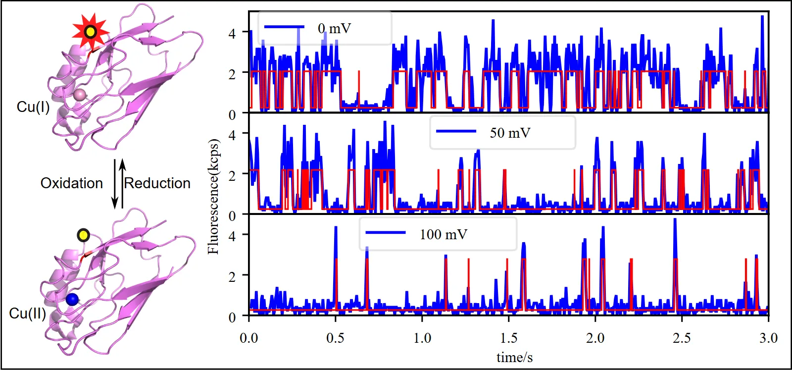
Azurin structure in the reduced Cu(I) and oxidised Cu(II) states, next to single-molecule fluorescence time traces at 0, 50 and 100 mV
Fig. 7A labelled azurin switches between Cu(I) and Cu(II); fluorescence traces at three redox potentials, with fitted states in red.
Chemical Science 11(3), 763–771 · 2020

Single electron transfer events and dynamical heterogeneity in the small protein azurin from Pseudomonas aeruginosa

Biswajit Pradhan, Christopher Engelhard, Sebastiaan Van Mulken, Xueyan Miao, Gerard W. Canters, Michel Orrit

Following the fluorescence of single dye-labelled azurin proteins, we timed individual electron-transfer events one by one. Their rates fluctuate over time even in this small 14 kDa protein, and comparing forward and backward reactions shows which molecular parameters cause the fluctuations.

Abstract

Monitoring the fluorescence of single-dye-labeled azurin molecules, we observed the reaction of azurin with hexacyanoferrate under controlled redox potential yielding data on the timing of individual (forward and backward) electron transfer (ET) events. Change-point analysis of the time traces demonstrates significant fluctuations of ET rates and of mid-point potential E0. These fluctuations are a signature of dynamical heterogeneity, here observed on a 14 kDa protein, the smallest to date. By correlating changes in forward and backward reaction rates we found that 6% of the observed change events could be explained by a change in midpoint potential, while for 25% a change of the donor–acceptor coupling could explain the data. The remaining 69% are driven by variations in complex association constants or structural changes that cause forward and back ET rates to vary independently. Thus, the observed spread in individual ET rates could be related in a unique way to variations in molecular parameters. The relevance for the understanding of metabolic processes is briefly discussed.

§ 02

All publications

2026

  1. SMC motor proteins operate at the near-minimal forces for DNA loop extrusion

    Adrian John Pinto, Biswajit Pradhan, Damla Tetiker, Maurice Pierre Schmitt, Eugene Kim, Peter Virnau

    bioRxiv (2026)preprint

    Abstract

    Loop extrusion by structural maintenance of chromosomes (SMC) complexes is essential for genome organization, yet the forces driving this process remain poorly understood. We present a coarse-grained model enabling predictive simulations of in vitro loop extrusion experiments at experimentally relevant time and length scales by matching parameters with concrete experiments. Using this model, we demonstrate that the extrusion forces generated by SMC motor proteins are just sufficient to overcome initial entropic barriers and sustain loop extrusion, highlighting that motors operate in the thermal regime. By measuring stalling tension directly, we confirm that they can be reliably determined by the Marko-Siggia equation and that varying grafting distances in experimental setups has only a marginal effect on the resulting tension. These results provide a predictive computation method for dissecting the mechanics of SMC driven genome folding.

2025

  1. Loop-extrusion-mediated plasmid DNA cleavage by the bacterial SMC Wadjet complex

    Biswajit Pradhan, Amar Deep, Jessica König, Martin D. Baaske, Kevin D. Corbett, Eugene Kim

    Molecular Cell 85(1), 107–116 (2025)

    Overview

    Bacteria defend themselves against invading plasmids with an SMC protein machine called Wadjet. Watching single Wadjet complexes at work, we found that they reel a plasmid's DNA into a loop from both sides at once, stall once the whole 44-kilobase plasmid has been pulled in, and then cut it. Loop extrusion is how Wadjet tells a plasmid apart and destroys it.

    Abstract

    Structural maintenance of chromosomes (SMC) complexes play pivotal roles in genome organization and maintenance across all domains of life. In prokaryotes, SMC-family Wadjet complexes structurally resemble the widespread MukBEF but serve a defensive role by inhibiting plasmid transformation. We previously showed that Wadjet specifically cleaves plasmid DNA; however, the molecular mechanism underlying plasmid recognition remains unclear. Here, we use in vitro single-molecule imaging to directly visualize DNA loop extrusion and plasmid cleavage by Wadjet. We find that Wadjet is a symmetric loop extruder that simultaneously reels in DNA from both sides of a loop and that this activity requires a dimeric JetABC supercomplex. On surface-anchored plasmid DNAs, Wadjet extrudes the full length of a 44-kb-pair plasmid, stalls, and cleaves DNA. Our findings reveal the role of loop extrusion in the specific recognition and elimination of plasmids by Wadjet and establish loop extrusion as an evolutionarily conserved mechanism among SMC complexes across all kingdoms of life.
2024

  1. Loop-extruding Smc5/6 organizes transcription-induced positive DNA supercoils

    Kristian Jeppsson, Biswajit Pradhan, Takashi Sutani, Toyonori Sakata, Miki Umeda Igarashi, Davide Giorgio Berta, Takaharu Kanno, Ryuichiro Nakato, Katsuhiko Shirahige, Eugene Kim, et al.

    Molecular Cell 84(5), 867–882 (2024) · cover article

    Overview

    In yeast chromosomes, Smc5/6 gathers where transcription over-twists DNA into positive supercoils. Single-molecule imaging shows why: Smc5/6 dimers recognise the tips of supercoiled DNA plectonemes, start extruding there, and collect the supercoiled DNA into one large plectonemic loop.

    Abstract

    The structural maintenance of chromosomes (SMC) protein complexes—cohesin, condensin, and the Smc5/6 complex (Smc5/6)—are essential for chromosome function. At the molecular level, these complexes fold DNA by loop extrusion. Accordingly, cohesin creates chromosome loops in interphase, and condensin compacts mitotic chromosomes. However, the role of Smc5/6's recently discovered DNA loop extrusion activity is unknown. Here, we uncover that Smc5/6 associates with transcription-induced positively supercoiled DNA at cohesin-dependent loop boundaries on budding yeast (Saccharomyces cerevisiae) chromosomes. Mechanistically, single-molecule imaging reveals that dimers of Smc5/6 specifically recognize the tip of positively supercoiled DNA plectonemes and efficiently initiate loop extrusion to gather the supercoiled DNA into a large plectonemic loop. Finally, Hi-C analysis shows that Smc5/6 links chromosomal regions containing transcription-induced positive supercoiling in cis. Altogether, our findings indicate that Smc5/6 controls the three-dimensional organization of chromosomes by recognizing and initiating loop extrusion on positively supercoiled DNA.

  2. Symmetry of loop extrusion by dimeric SMC complexes is DNA-tension-dependent

    Biswajit Pradhan, Adrian Pinto, Takaharu Kanno, Damla Tetiker, Martin D. Baaske, Erin Cutt, Constantinos Chatzicharlampous, Herwig Schüler, Amar Deep, Kevin D. Corbett, et al.

    bioRxiv (2024)preprint

    Overview

    SMC motors grow DNA loops by reeling in DNA from one side or from both. For the dimeric complexes Smc5/6 and Wadjet this turns out not to be fixed: at low DNA tension they pull from both sides, at higher tension they pull from one side at a time and can switch sides. Simulations show that thermal fluctuations drive this switching.

    Abstract

    Structural maintenance of chromosome (SMC) complexes organize and regulate genomes via DNA loop extrusion. During this process, the complexes increase the loop size by reeling in DNA from one or both sides of the loop. The factors governing this symmetry remain unclear. Here, we combine single-molecule analysis and molecular dynamic simulations to investigate the symmetry of loop extrusion of various SMC complexes. We find that whereas monomeric condensin and cohesin are one-sided extruders, the symmetry of dimeric SMCs, such as Smc5/6 and Wadjet, is DNA tension dependent. At low DNA tension (< 0.1pN), Smc5/6 and Wadjet extrude DNA from both sides of the loop. At higher tension, however, they transition to a behavior akin to one-sided extruders, yet still capable of extruding from one or the other side thereby switching the direction of extrusion. Our simulations further reveal that thermal fluctuations significantly influence loop extrusion symmetry, causing variations in DNA reeling rates between the two motors in the dimeric complexes and their direction switching at stalling tensions. Our findings challenge the previous view of loop extrusion symmetry as a fixed characteristic, revealing its dynamic nature and regulation by both intrinsic protein properties and extrinsic factors.

2023

  1. Testing pseudotopological and nontopological models for SMC-driven DNA loop extrusion against roadblock-traversal experiments

    Roman Barth†, Biswajit Pradhan†, Eugene Kim, Iain F. Davidson, Jaco van der Torre, Jan-Michael Peters, Cees Dekker

    Scientific Reports 13, 8100 (2023) · † equal contribution

  2. The Smc5/6 complex is a DNA loop-extruding motor

    Biswajit Pradhan, Takaharu Kanno, Miki Umeda Igarashi, Mun Siong Loke, Martin Dieter Baaske, Jan Siu Kei Wong, Kristian Jeppsson, Camilla Björkegren, Eugene Kim

    Nature 616, 843–848 (2023)

    Overview

    Cohesin and condensin were known to fold chromosomes by extruding DNA loops; what the third eukaryotic SMC complex, Smc5/6, does was unclear. Watching single molecules, we found that it is a loop-extruding motor too: as a dimer it reels DNA into loops from both sides at about one kilobase pair per second, while its Nse5/6 subunits act as a brake on starting new loops.

    Abstract

    Structural maintenance of chromosomes (SMC) protein complexes are essential for the spatial organization of chromosomes. Whereas cohesin and condensin organize chromosomes by extrusion of DNA loops, the molecular functions of the third eukaryotic SMC complex, Smc5/6, remain largely unknown. Using single-molecule imaging, we show that Smc5/6 forms DNA loops by extrusion. Upon ATP hydrolysis, Smc5/6 reels DNA symmetrically into loops at a force-dependent rate of one kilobase pair per second. Smc5/6 extrudes loops in the form of dimers, whereas monomeric Smc5/6 unidirectionally translocates along DNA. We also find that the subunits Nse5 and Nse6 (Nse5/6) act as negative regulators of loop extrusion. Nse5/6 inhibits loop-extrusion initiation by hindering Smc5/6 dimerization but has no influence on ongoing loop extrusion. Our findings reveal functions of Smc5/6 at the molecular level and establish DNA loop extrusion as a conserved mechanism among eukaryotic SMC complexes.

2022

  1. Condensin-driven loop extrusion on supercoiled DNA

    Eugene Kim, Alejandro Martin Gonzalez, Biswajit Pradhan, Jaco van der Torre, Cees Dekker

    Nature Structural & Molecular Biology 29(7), 719–727 (2022)

    Overview

    Chromosomal DNA in cells is twisted into supercoils. Imaging condensin on supercoiled DNA, we found that it prefers to bind near the tips of plectonemes and, as it extrudes a loop, collects the nearby supercoils into a single, highly stable supercoiled loop.

    Abstract

    Condensin, a structural maintenance of chromosomes (SMC) complex, has been shown to be a molecular motor protein that organizes chromosomes by extruding loops of DNA. In cells, such loop extrusion is challenged by many potential conflicts, for example, the torsional stresses that are generated by other DNA-processing enzymes. It has so far remained unclear how DNA supercoiling affects loop extrusion. Here, we use time-lapse single-molecule imaging to study condensin-driven DNA loop extrusion on supercoiled DNA. We find that condensin binding and DNA looping are stimulated by positively supercoiled DNA, and condensin preferentially binds near the tips of supercoiled plectonemes. Upon loop extrusion, condensin collects nearby plectonemes into a single supercoiled loop that is highly stable. Atomic force microscopy imaging shows that condensin generates supercoils in the presence of ATP. Our findings provide insight into the topology-regulated loading and formation of supercoiled loops by SMC complexes and clarify the interplay of loop extrusion and supercoiling.

  2. ParB proteins can bypass DNA-bound roadblocks via dimer-dimer recruitment

    Miloš Tišma, Maria Panoukidou, Hammam Antar, Young-Min Soh, Roman Barth, Biswajit Pradhan, Anders Barth, Jaco van der Torre, Davide Michieletto, Stephan Gruber, et al.

    Science Advances 8(26), eabn3299 (2022)

  3. SMC complexes can traverse physical roadblocks bigger than their ring size

    Biswajit Pradhan, Roman Barth, Eugene Kim, Iain F. Davidson, Benedikt Bauer, Theo van Laar, Wayne Yang, Je-Kyung Ryu, Jaco van der Torre, Jan-Michael Peters, et al.

    Cell Reports 41(3), 111491 (2022)

    Overview

    Chromosomal DNA is crowded with proteins, so how do ring-shaped SMC complexes extrude loops along it? Nucleosomes, RNA polymerase and dCas9 barely slow condensin down, and even 200-nm particles, far bigger than the SMC ring, are pulled into the loop. So the DNA does not thread through the ring, which points to a non-topological mechanism of loop extrusion.

    Abstract

    Ring-shaped structural maintenance of chromosomes (SMC) complexes like condensin and cohesin extrude loops of DNA. It remains, however, unclear how they can extrude DNA loops in chromatin that is bound with proteins. Here, we use in vitro single-molecule visualization to show that nucleosomes, RNA polymerase, and dCas9 pose virtually no barrier to loop extrusion by yeast condensin. We find that even DNA-bound nanoparticles as large as 200 nm, much bigger than the SMC ring size, also translocate into DNA loops during extrusion by condensin and cohesin. This even occurs for a single-chain version of cohesin in which the ring-forming subunits are covalently linked and cannot open to entrap DNA. The data show that SMC-driven loop extrusion has surprisingly little difficulty in accommodating large roadblocks into the loop. The findings also show that the extruded DNA does not pass through the SMC ring (pseudo)topologically, hence pointing to a nontopological mechanism for DNA loop extrusion.

2020

  1. Single electron transfer events and dynamical heterogeneity in the small protein azurin from Pseudomonas aeruginosa

    Biswajit Pradhan, Christopher Engelhard, Sebastiaan Van Mulken, Xueyan Miao, Gerard W. Canters, Michel Orrit

    Chemical Science 11(3), 763–771 (2020)

    Overview

    Following the fluorescence of single dye-labelled azurin proteins, we timed individual electron-transfer events one by one. Their rates fluctuate over time even in this small 14 kDa protein, and comparing forward and backward reactions shows which molecular parameters cause the fluctuations.

    Abstract

    Monitoring the fluorescence of single-dye-labeled azurin molecules, we observed the reaction of azurin with hexacyanoferrate under controlled redox potential yielding data on the timing of individual (forward and backward) electron transfer (ET) events. Change-point analysis of the time traces demonstrates significant fluctuations of ET rates and of mid-point potential E0. These fluctuations are a signature of dynamical heterogeneity, here observed on a 14 kDa protein, the smallest to date. By correlating changes in forward and backward reaction rates we found that 6% of the observed change events could be explained by a change in midpoint potential, while for 25% a change of the donor–acceptor coupling could explain the data. The remaining 69% are driven by variations in complex association constants or structural changes that cause forward and back ET rates to vary independently. Thus, the observed spread in individual ET rates could be related in a unique way to variations in molecular parameters. The relevance for the understanding of metabolic processes is briefly discussed.

2018

  1. Fluorescence of single copper proteins: dynamic disorder and enhancement by a gold nanorod

    Biswajit Pradhan

    PhD thesis, Leiden University (2018)thesis

    • Quantifying fluorescence enhancement for slowly diffusing single molecules in plasmonic near fields

      Martín Caldarola, Biswajit Pradhan, Michel Orrit

      The Journal of Chemical Physics 148(12) (2018)

    • Single-molecule fluorescence enhancement of a near-infrared dye by gold nanorods using DNA transient binding

      Weichun Zhang, Martín Caldarola, Xuxing Lu, Biswajit Pradhan, Michel Orrit

      Physical Chemistry Chemical Physics 20(31), 20468–20475 (2018)

    2017

    1. Gold nanorod enhanced fluorescence enables single-molecule electrochemistry of methylene blue

      Weichun Zhang, Martín Caldarola, Biswajit Pradhan, Michel Orrit

      Angewandte Chemie International Edition 56(13), 3566–3569 (2017)

    2016

    1. Gold-nanorod-enhanced fluorescence correlation spectroscopy of fluorophores with high quantum yield in lipid bilayers

      Biswajit Pradhan, Saumyakanti Khatua, Ankur Gupta, Thijs Aartsma, Gerard Canters, Michel Orrit

      The Journal of Physical Chemistry C 120(45), 25996–26003 (2016)

      Abstract

      Plasmonic fluorescence enhancement is used to study fluorescence correlation spectroscopy (FCS) at higher concentrations than in regular diffraction-limited FCS experiments. Previous studies suffered from sticking to the substrate and were performed mainly with poorly emitting dyes. A lipid bilayer forms a passivating surface, preventing sticking of the dye or the protein, and allows for specific anchoring of probe molecules. For dyes with high quantum yields, the fluorescence background of unenhanced molecules is high, and the fluorescence enhancement is weak, less than about 10. Nonetheless, we show that FCS is possible at micromolar concentrations of the probe molecule. Enhanced FCS is recorded by selecting signals on the basis of their shortened lifetime. This selection enhances the contrast of the correlation by more than an order of magnitude. The lipid bilayer can be used to anchor biomolecules and perform enhanced FCS, as we show for a dye-labeled protein.